What is Real Time Ultrasound Physiotherapy?
Real time ultrasound physiotherapy utilises an ultrasound machine to assist with the assessment and treatment of certain conditions. It provides real time imaging by transmitting sound waves through the body. These sound waves are then reflected by tissues in the body to create images on the screen for us to interpret.
The benefit of real time ultrasound physiotherapy is that it allows you to see how your muscles contract whilst performing certain exercises.
What Can Real Time Ultrasound Physiotherapy Be Used For?
Core Strengthening and Retraining
Often the cause of lower back pain can be related to poor core strength and stability. Whilst core strengthening exercises will help improve symptoms it can be hard to feel whether these deeply embedded muscles are switching on and therefore whether the exercises are being performed correctly. For this reason the core muscles are often assessed via real time ultrasound to give patients the opportunity to actually see these muscles working. These core muscles include the;
- transverse abdominis
- multifidus
- pelvic floor
- diaphragm
Together, these muscles act like a corset to provide spinal stability. Activating these muscles can be difficult as the movement is very small. Using real time ultrasound gives you instant feedback on how your muscles move and can be an extremely useful tool for retraining correct motor patterns.
The transverse abdominis is the deepest abdominal muscle and is initiated prior to movement. Many patients have difficulty activating this muscle correctly as pain can inhibit muscle activity. This can also occur to the multifidus.
The multifidus is a deep muscle in the back, also important for stability, especially with movements requiring spinal extension. Research shows that patients with back pain have delayed transverse abdominis and multifidus activation. Due to their role in spinal stability, it is no wonder why physiotherapists prescribe core exercises to the majority of patients suffering from back pain.
Another muscle that can assessed with real time ultrasound physiotherapy is the pelvic floor. This is often important for patients who suffer from incontinence and is generally assessed by a physiotherapist specialising in Womens’ Health (for more information visit Women in Focus Physiotherapy).
The diaphragm is not assessed on real time ultrasound but its role for spinal stability is not ignored. This is why patients are encouraged to maintain a normal breathing when performing the exercises prescribed. When patients have difficulty with an exercise they tend to hold their breath. This is a bracing strategy patients use to support themselves and can be retrained with correct cue’s and exercises to allow correct function of the diaphragm in stabilising the core.
Tendinopathy Assessment
In addition to assessing the core real time ultrasound physiotherapy can also be used in conjunction with physical assessments to assess tendinopathies. The most common tendons assessed on real time ultrasound include patella, achilles and gluteus medius tendons.
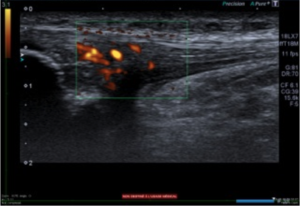
When a tendon is ill it can occasionally appear thickened on real time ultrasound. Hypoecoic (reflects relatively few ultrasound waves) areas are also visible which identifies that the tendon structure is altered. These areas show increased blood flow to the tendon on doppler imaging as shown below:
Real time ultrasound physiotherapy is an extremely useful treatment tool for a number of conditions and can assist patients in their rehabilitation. Patients often enjoy being able to see what is happening under their skin and can notice the improvements between sessions.
If you believe real time ultrasound physiotherapy could benefit you, or you wish to find out more about this treatment option, please contact our clinic by calling us on (02) 8323 7777 or email us at office@stadiumsportsphysio.com.au.
References
Bleakney, White and Maffulli (2007). The Achilles Tendon, Chapter 4- Imaging of the Achilles Tendon p25-38.
Kim, Cho, Goo and Baek (2013). Differences in Transverse Abdominis Muscle Function between Chronic Low Back Patients and Healthy Subjects at Maximum Expirations: Measurement with Real-time Ultrasonography. Journal of Physical Therapy Science. July 25(7): 861-863.
Peace, Lee and Healy (2006). Imaging the infrapatellar tendon in the elite athlete. Clinical Radiology 61, 570-578.